Description
Details
Description: Rabbit polyclonal antibody against the 2nd extracellular loop of human formyl peptide-receptor-like-1 (FPRL-1)
Purification: Protein G affinity purified
Product Type: Primary antibody
Target Protein: The 2nd extracellular loop of human formyl peptide-receptor-like-1 (FPRL1)
Immunogen: A short peptide (ASWGGTPEERLKC) corresponding to the amino acids on the 2nd extracellular loop of human FPRL1 is conjugated with KLH for immunization
Source: Rabbit
Specificity: This antibody is reactive to 38kDa human FPRL1.
Species Reacitvity: Human
Formulation: Lyophilized from a solution in 0.01M PBS pH7.2
Reconstitution: Double distilled water is recommended to reconstitute the antibody to 1mg/mL.
Storage: Store at -20oC. Avoid repeated freeze and thaw cycles.
Research Area: G-protein coupled membrane receptor, chemotaxis and Alzheimer’s disease
Background:
Human formyl peptide receptor-like 1(FPRL-1) is a G-protein coupled seven transmembrane receptor. The receptor can bind to the formyl peptides derived from the degradation of the cell wall of infected bacteria and host cells. FPRL1 expressed on the cell surface of neutrophils and other leukocytes mediates the chemotaxis of these cells toward infected site. It has been reported that acute phase protein SAA is also a ligand of FPRL1 and SAA can induce the secretion of IL-8 and TNF-α by neutrophil through FPRL1 signalling. FPRL1 has also been shown to mediate the chemotaxis of microglia by Abeta42 peptide and the internalization of Abeta42 peptide into cytoplasma of macrophage, indicating that FRPL1 may participate in the Abeta42 peptide elicited pathogenesis in Alzheimer’s disease.
Application:
1. Western Blot: Western blot analysis of HL-60 cell extract using 1:4000 to 1:5000 diluted anti-FPRL1 antibody.

2. Immunofluorescence: Analysis of HL-60 cells using 1:400 -1:500 diluted fluorescence labelled anti-FPRL1 antibody (green). Nuclei have been labelled with PI (red).

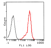
3. Flow Cytometry: The red lined peak (2nd peak) represents the histogram of HL-60 cells stained with 1:100 diluted anti- FPRL1 pAb. The black lined histogram (1st peak) represents negative control.
References:
If research is published using this product, please inform Anogen in order to cite the reference on this datasheet. Anogen will provide one unit of product in the same category as gratitude.
Additional
Additional Information
| Product Specificity | pAb Rabbit anti-Human FPRL-1 2nd Extracellular Loop |
|---|---|
| Application | WB, IF |
| Size | 0.1 mg |